
Antibody and Dye Based Assays

Biotium Histone H1(AE-4), CF488A conjugate, 0.1mg/mL
Eukaryotic histones are basic, water-soluble nuclear proteins that form hetero-octameric nucleosome particles. They wrap 146 base pairs of DNA in a left-handed supealpha-helicalturn sequentially to form chromosomal fiber. Two molecules of each of the four core histones (H2A, H2B, H3, and H4) form the octamer; formed of two H2A-H2B dimers and two H3-H4 dimers, forming two nearly symmetrical halves by tertiary structure. Over 80% of nucleosomes contain the linker Histone H1, derived from an intronless gene that interacts with linker DNA between nucleosomes and mediates compaction into higher order chromatin. Histones are subject to posttranslational modification by enzymes primarily on their N-terminal tails, but also in their globular domains. Such modifications include methylation, citrullination, acetylation, phosphorylation, sumoylation, ubiquitination and ADP-ribosylation.Primary antibodies are available purified, or with a selection of fluorescent CF Dyes and other lab

Biotium TAG-72 / CA72.4(CA72/145), CF488A conjugate, 0.1mg/mL
Recognizes an oncofetal antigen of 220 kDa, identified as a tumor-associated glycoprotein (TAG-72) with properties of a mucin. The majority of human adenocarcinomas including colorectal, pancreatic, gastric, ovarian, endometrial, mammary, and non-small cell lung cancer display some cell populations that are positive for TAG72. About 60% of carcinoma patients express TAG72 in their sera. TAG72 is reportedly useful in distinguishing pulmonary adenocarcinomas from pleural mesotheliomas.Primary antibodies are available purified, or with a selection of fluorescent CF Dyes and other labels. CF Dyes offer exceptional brightness and photostability. Note: Conjugates of blue fluorescent dyes like CF405S and CF405M are not recommended for detecting low abundance targets, because blue dyes have lower fluorescence and can give higher non-specific background than other dye colors.
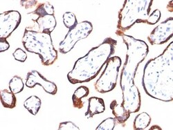
Biotium HCG-beta(HCGb/54), CF488A conjugate, 0.1mg/mL
This MAb reacts with a protein of 22 kDa, identified as β sub-unit of HCG. It does not cross react with the α sub-unit. HCG is a glycoprotein, which is secreted in large quantities by normal trophoblasts. It is present only in trace amounts in non-pregnant urine and sera but rises sharply during pregnancy. HCG is composed of two non-identical, non-covalently linked polypeptide chains designated as the alpha and beta subunits. The alpha subunit is identical to that of thyroid stimulating hormone (TSH), follicle stimulating hormone (FSH), and luteinizing hormone (LH). hCG MAb detects cells and tumors of trophoblastic origin such as choriocarcinoma. Large cell carcinoma and adenocarcinoma of the lung demonstrate anti-hCG positivity in 90% and 60% of cases respectively. 20% of lung squamous cell carcinomas are positive. hCG expression by non-trophoblastic tumors may indicate aggressive behavior.Primary antibodies are available purified, or with a selection of fluoresce
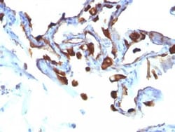
Biotium Cytokeratin 8/18(C-51), CF488A conjugate, 0.1mg/mL
Cytokeratin 8 (CK8) belongs to the type II (or B or basic) subfamily of high molecular weight cytokeratins and exists in combination with cytokeratin 18 (CK18). This MAb recognizes all simple epithelia including glandular epithelium, for example thyroid, female breast, gastrointestinal tract, respiratory tract, and urogenital tract including transitional epithelium. All adenocarcinomas and most squamous carcinomas are positive but keratinizing squamous carcinomas are usually negative. This antibody is useful in demonstrating the presence of Paget cells; there is very little keratin 18 in the normal epidermis so only Paget cells are stained.Primary antibodies are available purified, or with a selection of fluorescent CF Dyes and other labels. CF Dyes offer exceptional brightness and photostability. Note: Conjugates of blue fluorescent dyes like CF405S and CF405M are not recommended for detecting low abundance targets, because blue dyes have lower fluorescence and can give
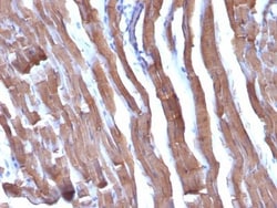
Biotium Liver Canuliculi(HSA98), CF488A conjugate, 0.1mg/mL
Monoclonal antibodies (MAbs) to liver cell processes are useful in the identification of hepatic carcinomas and normal organ structures. MAb HSA98 binds to human hepatocytes and the majority of human hepatocellular carcinomas (HCC's). In frozen sections, it stains hepatic cells and may be used as a marker of the liver. Cell preparations of hepatocellular carcinoma biopsies or cell lines are found to bind HSA98 on the cell surface. This MAb stains liver hepatocytes in frozen human liver sections and is positive on the cell surface of human liver carcinomas.Primary antibodies are available purified, or with a selection of fluorescent CF Dyes and other labels. CF Dyes offer exceptional brightness and photostability. Note: Conjugates of blue fluorescent dyes like CF405S and CF405M are not recommended for detecting low abundance targets, because blue dyes have lower fluorescence and can give higher non-specific background than other dye colors.
Biotium MART-1 / Melan-A(M2-9E3), CF488A conjugate, 0.1mg/mL
This antibody recognizes a protein doublet of 20-22 kDa, identified as MART-1 (Melanoma Antigen Recognized by T cells 1) or Melan-A. MART-1 is a newly identified melanocyte differentiation antigen recognized by autologous cytotoxic T lymphocytes. Seven other melanoma associated antigens recognized by autologous cytotoxic T cells include MAGE-1, MAGE-3, tyrosinase, gp100, gp75, BAGE-1, and GAGE-1. Subcellular fractionation shows that MART-1 is present in melanosomes and endoplasmic reticulum. This MAb labels melanomas and other tumors showing melanocytic differentiation. It is also a useful positive-marker for angiomyolipomas. It does not stain tumor cells of epithelial, lymphoid, glial, or mesenchymal origin.Primary antibodies are available purified, or with a selection of fluorescent CF Dyes and other labels. CF Dyes offer exceptional brightness and photostability. Note: Conjugates of blue fluorescent dyes like CF405S and CF405M are not recommended for detecting low abu
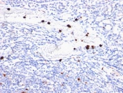
Biotium Granulocyte Marker(BM-2), CF488A conjugate, 0.1mg/mL
Recognizes 183 kDa protein with DNA-binding characteristics, which is identified as a myeloid specific antigen. It reacts with myeloid precursor cells and granulocytes in bone marrow. Its antigen appears to be restricted to M2 and M3 acute myelogenous leukemia (AML) subtypes. Markers of myeloid cells are useful in the identification of different levels of cellular differentiation. This MAb reacts with early precursor and mature forms of human myeloid cells. It is useful in the identification of myelogenous leukemias, distinguishing granulocytic sarcomas from lymphoid malignancies and also in the study of differentiation and transformation of human myeloid cells. The biological function of this antigen is not clear, although it has been proposed that it may play a role in the differentiation of myeloid cells.Primary antibodies are available purified, or with a selection of fluorescent CF Dyes and other labels. CF Dyes offer exceptional brightness and photostability. Note: C
Biotium CD56 / NCAM(123C3.D5), CF488A conjugate, 0.1mg/mL
This MAb reacts with an extracellular domain (close to transmembrane) of CD56/NCAM. Three isoforms of neural cell adhesion molecule (NCAM) are produced by differential splicing of the RNA transcript from a single gene. The 135 kDa isoform is the basic molecule, which is glycosylated or sialylated to produce the mature species. Anti-CD56 recognizes two proteins of the neural cell adhesion molecule, the basic molecule expressed on most neuroectodermally derived tissues and neoplasms (e.g. retinoblastoma, medulloblastomas, astrocytomas, neuroblastomas, and small cell carcinomas). It is also expressed on some mesodermally derived tumors (rhabdomyosarcoma). Anti-CD56 plays an important role in the diagnosis of nodal and nasal NK/T-cell lymphomas.Primary antibodies are available purified, or with a selection of fluorescent CF Dyes and other labels. CF Dyes offer exceptional brightness and photostability. Note: Conjugates of blue fluorescent dyes like CF405S and CF405M are
Biotium MART-1 / Melan-A(M2-7C10), CF488A conjugate, 0.1mg/mL
This antibody recognizes a protein doublet of 20-22 kDa, identified as MART-1 (Melanoma Antigen Recognized by T cells 1) or Melan-A. MART-1 is a newly identified melanocyte differentiation antigen recognized by autologous cytotoxic T lymphocytes. Seven other melanoma associated antigens recognized by autologous cytotoxic T cells include MAGE-1, MAGE-3, tyrosinase, gp100, gp75, BAGE-1, and GAGE-1. Subcellular fractionation shows that MART-1 is present in melanosomes and endoplasmic reticulum. This MAb labels melanomas and other tumors showing melanocytic differentiation. It is also a useful positive-marker for angiomyolipomas. It does not stain tumor cells of epithelial, lymphoid, glial, or mesenchymal origin.Primary antibodies are available purified, or with a selection of fluorescent CF Dyes and other labels. CF Dyes offer exceptional brightness and photostability. Note: Conjugates of blue fluorescent dyes like CF405S and CF405M are not recommended for detecting low abu
Biotium CD20(109-3C2), CF488A conjugate, 0.1mg/mL
Recognizes a protein of 30-33 kDa, which is identified as CD20 (Workshop V; Code CD20.12. Workshop IV; Code B17). It recognizes an extracellular domain of CD20. It is a non-Ig differentiation antigen of B-cells and its expression is restricted to normal and neoplastic B-cells, being absent from all other leukocytes and tissues. CD20 is expressed by pre B-cells and persists during all stages of B-cell maturation but is lost upon terminal differentiation into plasma cells. The protein passes through the membrane 4 times with both ends in cytoplasm and exposes one short and one longer loop to the external environment. CD20 is not glycosylated in resting B-cells and its cytoplasmic domains are differentially phosphorylated upon activation. It acts as calcium channel involved in B cell activation and cell cycle progression.Primary antibodies are available purified, or with a selection of fluorescent CF Dyes and other labels. CF Dyes offer exceptional brightness and photostabilit
Biotium IGF-1(M23), CF488A conjugate, 0.1mg/mL
This antibody is specific to Insulin-like Growth Factor (IGF-1) and shows minimal cross-reaction with IGF-11, Proinsulin, MSF, and Insulin. IGF-1 is a polypeptide growth factor with two isoforms that are produced by alternative splicing. Isoform 1 is also known as IGF-IB while isoform 2 is known as IGF-IA. IGF-1 stimulates the proliferation of a wide range of cell types including muscle, bone and cartilage tissue. It functions as an autocrine regulator of growth. Activation of IGF system has emerged as a key factor for tumor progression and resistance to apoptosis in many cancers like those of breast, thyroid and colon.Primary antibodies are available purified, or with a selection of fluorescent CF Dyes and other labels. CF Dyes offer exceptional brightness and photostability. Note: Conjugates of blue fluorescent dyes like CF405S and CF405M are not recommended for detecting low abundance targets, because blue dyes have lower fluorescence and can give higher non-specific
Biotium CD56 / NCAM(NCAM1/795), CF488A conjugate, 0.1mg/mL
This MAb reacts with an extracellular domain (close to transmembrane) of CD56/NCAM. Three isoforms of neural cell adhesion molecule (NCAM) are produced by differential splicing of the RNA transcript from a single gene. The 135 kDa isoform is the basic molecule, which is glycosylated or sialylated to produce the mature species. Anti-CD56 recognizes two proteins of the neural cell adhesion molecule, the basic molecule expressed on most neuroectodermally derived tissues and neoplasms (e.g. retinoblastoma, medulloblastomas, astrocytomas, neuroblastomas, and small cell carcinomas). It is also expressed on some mesodermally derived tumors (rhabdomyosarcoma). Anti-CD56 plays an important role in the diagnosis of nodal and nasal NK/T-cell lymphomas.Primary antibodies are available purified, or with a selection of fluorescent CF Dyes and other labels. CF Dyes offer exceptional brightness and photostability. Note: Conjugates of blue fluorescent dyes like CF405S and CF405M are not
Biotium Glypican-3(GPC3/863 + 1G12), CF488A conjugate, 0.1mg/mL
Glypican-3 (GPC3) is an integral membrane protein that is mutated in the Simpson-Golabi-Behmel syndrome (SGBS). SGBS is characterized by pre- and post-natal overgrowth and is a recessive X-linked condition. GPC3 may also be found in a secreted form. Anti-GPC3 has been identified as a useful tumor marker for the diagnosis of hepatocellular carcinoma (HCC), hepatoblastoma, melanoma, testicular germ cell tumors, and Wilm's tumor. In patients with HCC, GPC3 is overexpressed in neoplastic liver tissue and elevated in serum, but is undetectable in normal liver, benign liver, and the serum of healthy donors. GPC3 expression is also found to be higher in HCC liver tissue than in cirrhotic liver or liver with focal lesions such as dysplastic nodules and areas of hepatic adenoma (HA) with malignant transformation. In the context of testicular germ cell tumors, GPC3 expression is up regulated in certain histologic subtypes, specifically yolk sac tumors and choriocarcinoma. A high level